
Building a healthier bottomline, together
Find solutions to your biggest challenges


Insights and trends in healthcare
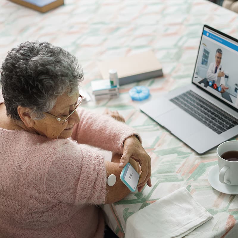
Futurizing healthcare experiences to drive ROI Articles
ArticlesFuturizing healthcare experiences to drive ROI
5 strategies healthcare organizations can implement to create better patient and provider experiences.
Reimagining how we care for healthcare workers Industry report
Industry reportReimagining how we care for healthcare workers
Get the latest insights on accelerating technology adoption to reduce talent strain.
Ricoh 3D anatomic models gain FDA 510(k) clearance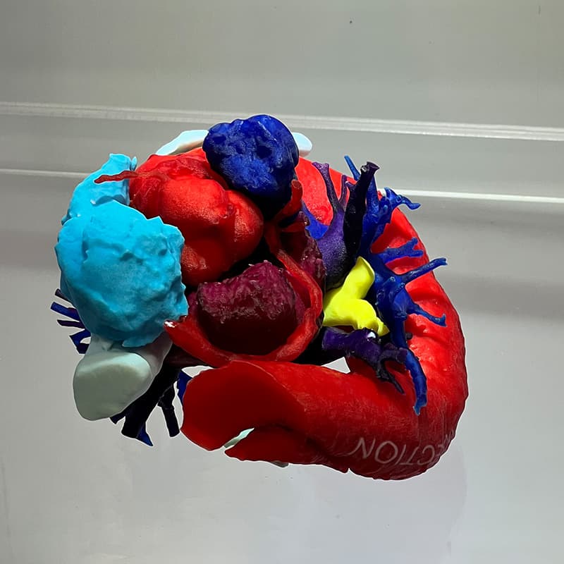 Articles
ArticlesRicoh 3D anatomic models gain FDA 510(k) clearance
Ricoh’s FDA 510(k) clearance means healthcare providers don’t have to be experts in printing 3D models.


Achieving real customer results in healthcare
Solve claims processing delays
Automating the flow of information through your organization reduces the risk of common manual errors and increases the speed of review, approvals, and patient response.

Elevate the patient experience
Improve appointment preparation and patient recovery times with customized packet and letter automation, ensuring information is communicated correctly and promptly while freeing staff resources and reducing business costs.

Speak with a representative
- 1. FDA-cleared medical device for diagnostic use within craniomaxillofacial (CMF), orthopedic, cardiovascular, neurological, gastrointestinal, genitourinary, and breast applications.